
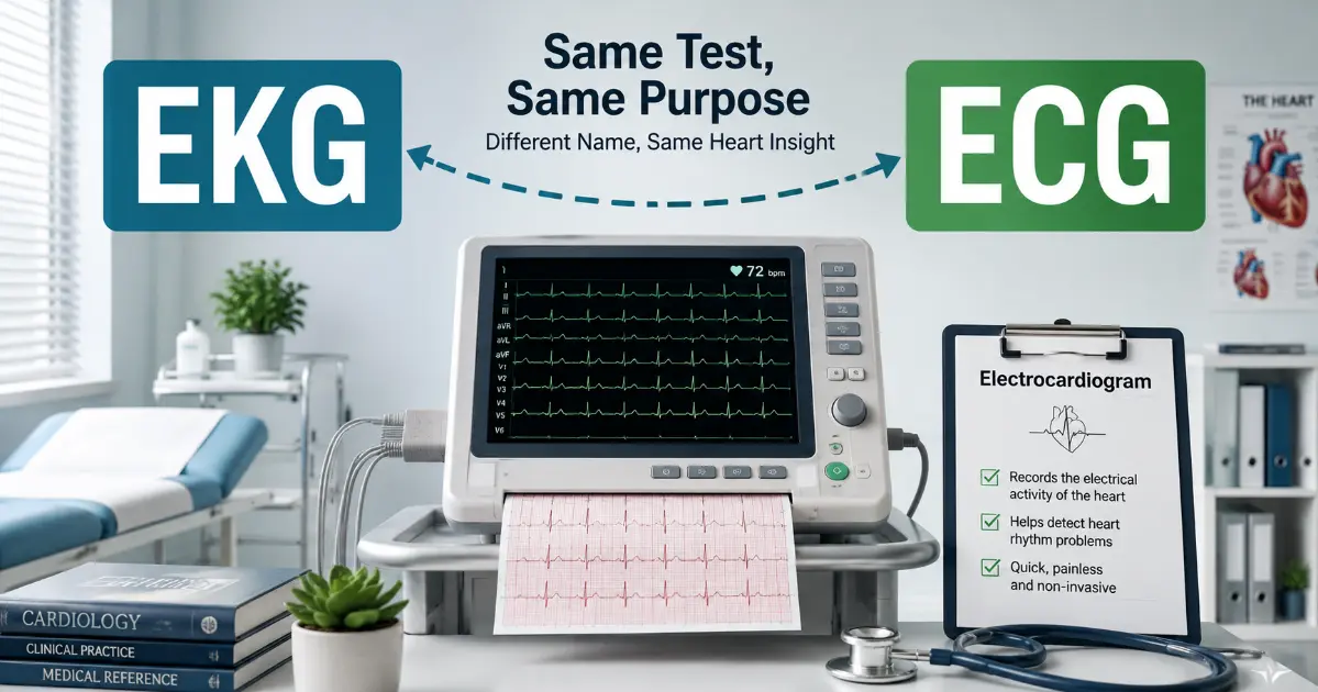
If you’ve searched for ekg vs ecg, you’re probably wondering if these are two different heart tests. They aren’t. EKG and ECG are two abbreviations for the same test called an electrocardiogram. The test records the electrical activity of your heart and helps health care providers check your heart rate and rhythm.
The only real difference is the abbreviation. ECG comes from the English spelling electrocardiogram. EKG comes from the German spelling Elektrokardiogramm, which uses a K. Both terms refer to the same test and the same basic procedure.
You may see EKG on a medical form and ECG on a health website. That doesn’t mean you’re getting different tests. The machine and the basic procedure are the same. The different letters simply come from different versions of the word.
This guide explains why both abbreviations exist, what an electrocardiogram measures, how the test works, what the results can show, and how it differs from other heart tests. You’ll also learn what terms such as 12 lead ECG, resting ECG, and stress test mean.
Quick Definition
An ECG and an EKG are the same medical test. Both abbreviations refer to an electrocardiogram, which records the electrical activity of the heart.
The test can show your heart rate and rhythm and can provide clues about how electrical signals move through your heart. Doctors may use it to look for signs of heart rhythm problems, heart damage, and other heart conditions.
Direct Answer
EKG and ECG mean the same thing. There is no difference in the test itself. EKG comes from the German spelling of electrocardiogram, while ECG follows the English spelling.
Comparison Table
| Term | Meaning | Example | Common Use |
|---|---|---|---|
| ECG | Electrocardiogram | The doctor ordered an ECG to check the heart rhythm. | Common in medical writing and many countries. |
| EKG | Electrocardiogram | The nurse performed an EKG before surgery. | Common in the United States and clinical settings. |
| Electrocardiogram | Full name of the test | The electrocardiogram records the heart’s electrical activity. | Formal medical language and patient information. |
Why There Are Two Names
The two abbreviations come from the same medical term. The difference comes from language rather than medicine.
In English, the test is called an electrocardiogram. The first letters give us ECG. The abbreviation follows the English spelling of the word.
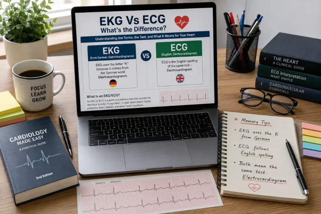
The German name is Elektrokardiogramm. It uses a K in the middle of the word, so the abbreviation becomes EKG. The K reflects the German spelling and does not indicate a different type of test.
This can seem strange at first. After all, English uses the letter C in cardiogram, so why would a medical test in the United States use K? The answer goes back to the history of the term and the influence of German medical language.
Why The K Appears In EKG
The K comes from the German word Kardiogramm. German uses K where English uses C in this part of the word.
The abbreviation stayed in medical use even after the English form became common. That is why you can find EKG and ECG used side by side today.
The K does not stand for a special heart signal. It does not mean the test uses a different machine. It does not indicate a different number of electrodes or a different type of recording.
Think of EKG and ECG as two labels for the same thing. The spelling changes, but the test does not.
Why EKG Is Common In The United States
You’ll often see EKG in American hospitals, clinics, doctors’ offices, and patient records. It has become a familiar term in US health care.
At the same time, ECG is also widely used in the United States. You may see it in medical articles, research, educational materials, and health information.
This means you shouldn’t worry if one doctor calls it an EKG while another calls it an ECG. They are talking about the same heart test.
The same applies when reading medical information online. If one page explains ECG results and another talks about EKG results, you don’t need to learn two separate procedures. Both terms describe an electrocardiogram.
What An EKG Or ECG Measures
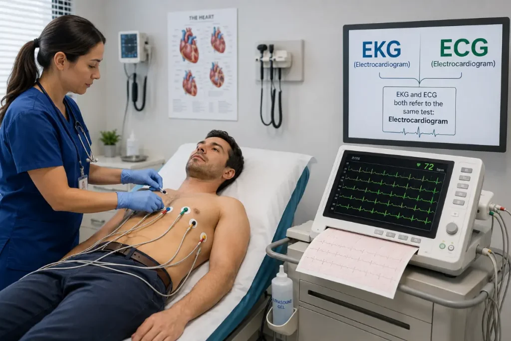
An electrocardiogram records the electrical activity of your heart. Every heartbeat depends on an electrical signal that tells the heart muscle when to contract.
The signal normally begins in a part of the heart called the sinoatrial node, or SA node. It then moves through the heart in a controlled pattern. This electrical process helps the upper and lower chambers contract at the right times.
The electrodes placed on your skin detect these electrical signals. Wires carry the information to the machine, which creates a tracing on a screen or a printed page.
That tracing gives a health care provider several useful pieces of information. It can show how quickly your heart is beating, if the rhythm appears regular, and how the electrical signal moves through different parts of the heart.
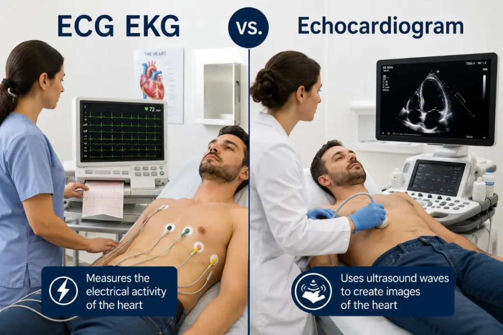
The test doesn’t take a picture of your heart. It records electrical activity instead. That makes it different from tests such as an echocardiogram, which uses sound waves to create images of the heart.
The Heart’s Electrical System
Your heart needs a well organized electrical system to beat properly. The electrical signal normally starts in the SA node in the upper part of the heart.
From there, the signal travels through the upper chambers. It then moves toward the lower chambers, causing them to contract.
This timing matters. The upper chambers help fill the lower chambers with blood. The lower chambers then pump blood to the lungs and the rest of the body.
An electrocardiogram records the electrical changes linked to this process. A health care provider can study the pattern to see if the heartbeat appears normal or if there are signs that something needs more attention.
What The Waves Mean
If you look at an ECG tracing, you’ll see several waves and sections. Three important parts are the P wave, QRS complex, and T wave.
The P wave represents the electrical activity linked to the upper chambers of the heart. The QRS complex represents the electrical activity linked to the lower chambers. The T wave represents the recovery phase of the lower chambers after they contract.
You don’t need to memorize these terms to understand the basic purpose of the test. They are mainly useful because health care professionals use the shape, timing, and spacing of these parts to assess heart activity.
For example, a provider may look at the distance between certain waves to see how quickly electrical signals are moving through the heart. Changes in the pattern can provide clues about heart rhythm or electrical conduction.
An abnormal tracing does not automatically mean you have a serious heart problem. The result needs to be considered alongside your symptoms, medical history, physical exam, and other test results.
How The Test Works
A standard electrocardiogram is quick and noninvasive. Noninvasive means that the test doesn’t require anything to be inserted into your body.
You normally lie down while a health care professional places small adhesive electrodes on your skin. The electrodes are usually placed on your chest and limbs. Wires connect them to the recording machine.
Once everything is connected, you’ll need to stay still for a short time. The machine records your heart’s electrical activity and produces a tracing.
The test itself is usually very quick. The preparation can take longer than the actual recording because the health care professional needs to position the electrodes correctly.
The machine doesn’t send electricity into your body during a standard test. It simply detects the electrical signals produced by your heart.
What The Electrodes Do
The small sticky patches used during the test are called electrodes. Their job is to detect electrical activity from your heart through your skin.
The electrodes don’t make your heart beat. They don’t shock your heart. They simply pick up the electrical signals that are already there.
Wires connect the electrodes to the recording machine. The machine uses those signals to create the lines and waves you see on the ECG tracing.
You may notice several electrodes placed around your chest and on your arms or legs. Their positions allow the machine to record the heart’s electrical activity from different angles.
What A 12 Lead Test Means
You may hear the phrase 12 lead ECG or 12 lead EKG when discussing heart testing.
The word lead can be confusing because it sounds like it refers to a wire. In this context, a lead is an electrical view of the heart.
A standard 12 lead test creates 12 different views of the heart’s electrical activity. These views help a health care provider examine the heart from different angles.
The test uses electrodes placed on specific areas of the body to produce these views. So the phrase 12 lead does not simply mean that twelve stickers are placed on your skin.
This is one reason the electrode placement matters. Moving an electrode to the wrong position can affect the tracing and make it harder to interpret accurately.
Does The Test Hurt
A standard electrocardiogram is generally painless. You may feel a little discomfort when the adhesive electrodes are placed or removed.
Some people have sensitive skin and may notice mild redness or irritation after the electrodes come off. This usually goes away on its own.
The test doesn’t involve radiation. It also doesn’t send electricity through your body.
If you have body hair where the electrodes need to be placed, a health care professional may need to remove a small amount of hair so the electrodes can stick properly. That can be mildly uncomfortable, but the recording itself shouldn’t hurt.
How Long Does An EKG Take
A standard resting test usually takes only a few minutes from start to finish. The exact time depends on how quickly the electrodes can be placed and how much preparation is needed.
The actual recording can be very short. In many cases, the machine needs only a few seconds to capture the heart’s electrical pattern.
Longer testing is different. An exercise stress test, for example, involves recording your heart activity while you exercise. A portable monitor may record your heart rhythm for many hours or even several days.
So when someone says they are having an EKG, the length of the appointment can depend on the type of heart monitoring being used.
Why A Doctor May Order The Test
Doctors use electrocardiograms for many different reasons. The test can help investigate symptoms and provide information about how the heart is working.
A doctor may order one if you have chest discomfort, a fast heartbeat, an irregular heartbeat, dizziness, fainting, shortness of breath, or unusual tiredness. The test may also be used if you have a known heart condition or need your heart monitored during treatment.
An electrocardiogram may also be part of a medical evaluation before surgery. The decision to order one depends on your health, symptoms, age, medical history, and the type of procedure you are having.
Doctors can also use ECG testing to monitor certain medications or medical devices. In some situations, repeated tests can help show how the heart changes over time.
The test provides useful information quickly, but it doesn’t answer every question about the heart. A normal result doesn’t rule out every possible heart condition. An abnormal result doesn’t automatically mean that you have a serious disease.
Your health care provider interprets the result in context. They may compare it with an older tracing, review your symptoms, or order another test if they need more information.
That is an important point to remember. An ECG or EKG is a valuable tool, but it is only one part of a complete heart evaluation.
What An EKG Can Show
An electrocardiogram can give a health care provider a quick look at the heart’s electrical activity. The tracing can reveal patterns that may suggest an irregular rhythm, a problem with electrical conduction, or signs of reduced blood flow to the heart.
The test can also show the heart rate. A heart that beats too quickly is called tachycardia. A heart that beats too slowly is called bradycardia. An ECG can help identify these patterns, although the meaning of the result depends on the person and the situation.
The rhythm is another major part of the reading. A healthy heart usually follows a regular electrical pattern. Some rhythm problems cause the heartbeat to become irregular or unusually fast. An ECG can capture these changes when they occur during the recording.
Heart Rate
Your heart rate tells you how many times your heart beats in a minute. An ECG can measure this rate very accurately during the test.
A resting adult heart rate often falls between 60 and 100 beats per minute. That range is a general guide rather than a rule that applies to everyone. Athletes, for example, can have lower resting heart rates without having a heart problem.
The number alone doesn’t tell the whole story. A doctor also looks at the rhythm and the pattern of the electrical signals.
For example, a person might have a heart rate of 90 beats per minute with a regular rhythm. Another person might have the same rate but an irregular rhythm. Those two tracings can mean very different things.
Heart Rhythm
Heart rhythm describes how regularly the heart beats. An ECG can help identify many types of irregular rhythms.
Atrial fibrillation is one well known example. It can cause an irregular heartbeat because the upper chambers of the heart don’t follow their usual coordinated electrical pattern.
Other rhythm problems can cause the heart to beat very quickly or very slowly. Some can come and go, which means a short ECG might not catch them.
That is one reason doctors sometimes use longer forms of heart monitoring. A portable monitor can record heart activity for a much longer period and may catch an irregular rhythm that doesn’t happen during a short office test.
Electrical Conduction
The heart relies on electrical signals moving through specific pathways. These signals help different parts of the heart contract in the right order.
An ECG can show how quickly these signals move through the heart. A delay or unusual pattern can point to a conduction problem.
Doctors may look closely at the timing between different parts of the tracing. These measurements can provide clues about how the electrical system is working.
This doesn’t mean that every unusual measurement points to a disease. Some variations can be normal for a particular person. Doctors consider the complete tracing along with the person’s symptoms and medical history.
Signs Of A Heart Attack
An ECG can provide important information when a heart attack is suspected. Certain changes in the electrical pattern can suggest that part of the heart muscle isn’t receiving enough blood.
Doctors may perform an ECG when someone arrives with symptoms such as chest pressure, shortness of breath, sweating, nausea, or pain that spreads to the arm, back, neck, or jaw.
The test is especially useful because it can be performed quickly. In an emergency, doctors can use the result along with symptoms and other tests to decide what needs to happen next.
An ECG cannot detect every heart attack by itself. Some heart attacks may not produce obvious changes on the first recording. Doctors may repeat the test or use blood tests and other examinations when needed.
Signs Of Previous Heart Damage
An ECG can sometimes show patterns that suggest the heart has been damaged in the past. For example, certain changes may be consistent with an old heart attack.
The tracing doesn’t always tell a doctor exactly when an injury happened. It also doesn’t provide a complete picture of the heart’s structure.
That is why an ECG result is usually interpreted together with other information. A doctor may compare the new tracing with an older one to see if anything has changed.
EKG Vs ECG For Different Situations
The name of the test doesn’t change based on the reason it is performed. An ECG and an EKG are still the same basic test.
What can change is the type of monitoring used. A person might have a standard resting ECG in a doctor’s office. Another person might have an exercise test or wear a portable monitor at home.
These tests all examine electrical activity in some way, but they answer different questions.
Resting ECG
A resting ECG records your heart while you are lying or sitting quietly. It is one of the most common forms of electrocardiogram testing.
The test gives a snapshot of your heart’s electrical activity at that moment. It can be useful for checking symptoms, identifying rhythm problems, and looking for certain signs of heart disease.
Because the recording is short, it may not catch a problem that happens only occasionally. Someone who has episodes of a racing or irregular heartbeat might have a normal resting ECG if the symptoms aren’t happening during the test.
Exercise ECG
An exercise ECG records the heart while you are physically active. You may walk on a treadmill or use a stationary bicycle while medical staff monitor your heart.
Exercise makes the heart work harder. This can reveal changes that aren’t visible while you are resting.
Doctors may use an exercise test to evaluate symptoms that appear during physical activity. It can also provide information about how the heart responds to increased demand.
The test is performed under medical supervision. The health care team watches your heart rate, rhythm, blood pressure, and symptoms throughout the procedure.
Portable Heart Monitoring
Some heart rhythm problems happen only once in a while. A short office ECG might miss them.
A portable monitor can help in these cases. Depending on the device, you may wear it for a day, several days, or longer.
A Holter monitor is one example. It continuously records heart activity while you go about your normal routine.
Another type of device may record only when symptoms occur or when you activate it. Your doctor will choose the type of monitor based on your symptoms and how often they happen.
How An ECG Differs From An Echocardiogram
The names sound similar, so people often confuse an electrocardiogram with an echocardiogram. They are not the same test.
An ECG records electrical activity. An echocardiogram uses sound waves to create moving images of the heart.
Think of an ECG as a record of the heart’s electrical signals. An echocardiogram is more like a live picture of the heart.
An echocardiogram can show the size and shape of the heart and how its chambers and valves are working. It can also show how well the heart muscle moves.
An ECG answers a different set of questions. It focuses on heart rate, rhythm, and electrical activity.
Doctors sometimes use both tests because they provide different information. One test cannot replace the other.
ECG And Heart Ultrasound
An echocardiogram is often called a heart ultrasound. It uses sound waves rather than electrical sensors to create images.
During the test, a technician moves an ultrasound device over the chest. The machine creates images that show the heart as it moves.
This can help doctors examine the heart valves, chambers, muscle, and pumping function. It can also help identify structural problems that an ECG cannot show.
The two tests can work well together. One looks at electrical activity while the other looks at the heart’s physical structure and movement.
How An ECG Differs From A Heart Monitor
A standard ECG is usually a short recording. A heart monitor can record electrical activity for much longer.
This difference matters when symptoms come and go.
Imagine someone who feels their heart racing once every few days. If they have a short ECG at the doctor’s office when they feel normal, the result may not capture the episode.
A portable monitor can record heart activity during daily life. If the irregular rhythm happens while the person is wearing the device, the recording may capture it.
The choice depends on the symptoms. A doctor may start with a standard ECG and then recommend longer monitoring if the situation calls for it.
Understanding Common ECG Terms
Medical reports can look confusing because they contain technical words and abbreviations. Learning a few common terms can make them easier to understand.
Sinus Rhythm
Sinus rhythm describes a heartbeat that starts from the heart’s normal natural pacemaker. The SA node normally creates the electrical signal that starts each heartbeat.
A report may describe a tracing as normal sinus rhythm. That generally means the electrical rhythm follows the expected pattern from the heart’s natural pacemaker.
The phrase doesn’t mean every part of the heart is guaranteed to be healthy. It describes a specific part of the heart’s electrical activity.
Arrhythmia
An arrhythmia is an abnormal heart rhythm. The heart may beat too quickly, too slowly, or irregularly.
Some arrhythmias are harmless. Others can require treatment or further testing.
An ECG can help identify many rhythm problems. Still, some arrhythmias happen only occasionally and may require longer monitoring to detect.
Heart Rate
Heart rate is the number of heartbeats per minute. An ECG can measure the rate during the recording.
A fast or slow rate doesn’t automatically mean something is wrong. Exercise, stress, medications, sleep, fitness level, and other factors can affect heart rate.
Doctors look at the number along with the rhythm and the person’s overall health.
QRS Complex
The QRS complex is one of the main parts of an ECG tracing. It represents the electrical activity associated with the lower chambers of the heart.
Doctors examine its shape and timing as part of the overall ECG reading.
A person doesn’t need to understand every detail of the QRS complex to know what their test means. If a report mentions an unusual QRS pattern, the health care provider can explain what it means in that specific case.
PR Interval
The PR interval measures the time between the start of electrical activity in the upper chambers and the start of electrical activity in the lower chambers.
It provides information about how the electrical signal travels through the heart.
Doctors use this measurement as part of a larger assessment. One number rarely tells the complete story.
QT Interval
The QT interval represents a period of electrical activity and recovery in the heart’s lower chambers.
Doctors pay attention to the QT interval because certain medications, medical conditions, and electrolyte problems can affect it.
A report may list a corrected QT value because heart rate can affect the length of the QT interval. Your health care provider can explain the result based on your specific situation.
Why ECG Results Can Look Scary
Looking at an ECG tracing for the first time can be intimidating. The page may contain sharp peaks, curved waves, numbers, and medical abbreviations that seem impossible to understand.
Remember that an ECG is designed for trained professionals to interpret. The lines are not meant to be read like ordinary text.
An unusual looking wave doesn’t automatically mean something is wrong. The interpretation depends on the full pattern, the person’s age, symptoms, health history, and other findings.
The same is true for numbers on an ECG report. A value outside a commonly listed range doesn’t automatically mean a serious problem.
If you receive an ECG report and don’t understand a term, ask the health care professional who ordered the test. They can explain what the finding means in the context of your health.
Common Misunderstandings About EKG And ECG
The difference in spelling creates several common misunderstandings. Most are easy to clear up once you know that both terms refer to the same test.
They Are Not Two Different Tests
This is the biggest misunderstanding.
An EKG isn’t a different version of an ECG. The two names refer to the same electrocardiogram.
If your doctor orders an EKG and you later see ECG on your medical records, you don’t need to assume another test was performed.
EKG Does Not Mean A Stronger Test
Some people think the K makes EKG a special or more detailed version. It doesn’t.
The abbreviation has nothing to do with how advanced the test is. It comes from the German spelling of the medical term.
A basic EKG and a basic ECG are the same type of test.
ECG Does Not Mean A Different Machine
The same misunderstanding can happen in reverse. Seeing ECG instead of EKG doesn’t mean the hospital is using a different machine.
The equipment may look different from one clinic to another, but that has nothing to do with the abbreviation.
The name simply depends on the terminology the medical provider chooses to use.
Which Term Should You Use
If you’re writing for a US audience, EKG is a very familiar term. Many patients recognize it immediately because they have heard doctors and nurses use it.
ECG is also correct and widely understood. It is especially common in international medical writing and scientific discussions.
If you’re writing an article for general readers, you can introduce both terms at the beginning. For example, you can write that an electrocardiogram is also called an ECG or EKG. After that, choose one term and use it consistently.
This keeps your writing easy to follow. It also prevents readers from thinking you are talking about two different tests.
If you’re speaking with a health care provider, you can use either abbreviation. The provider will understand what you mean.
How To Remember The Difference
The easiest memory trick is to focus on the word behind the abbreviation.
ECG follows the English word electrocardiogram.
EKG follows the German word Elektrokardiogramm.
The K comes from the German spelling. That’s the whole difference.
You don’t need to remember two sets of medical facts. You only need to remember that the letters come from different languages.
A simple way to think about it is this.
C means English spelling.
K means German spelling.
Both lead to the same heart test.
When An ECG May Be Normal
A normal ECG is generally a reassuring finding, but it doesn’t mean every possible heart problem has been ruled out. An electrocardiogram records your heart’s electrical activity during the time of the test.
That matters because some heart problems happen only at certain times. A person may have symptoms at home but feel completely normal during a short appointment. In that situation, the ECG may look normal even though the symptoms still deserve attention.
Doctors consider the test alongside your symptoms, health history, physical exam, and other results. They may also compare the tracing with an older ECG to look for changes.
A Normal Result Is Only One Piece Of The Picture
It is easy to think of a normal test as a complete health check. It isn’t.
An ECG can answer useful questions about your heart rhythm and electrical activity. It cannot show every problem involving the heart muscle, valves, blood vessels, or other structures.
For example, an ECG may not detect a problem that happens only during exercise if you’re resting during the recording. Your doctor may recommend another type of test if your symptoms suggest that more information is needed.
This is why you shouldn’t judge a heart problem based on one number or one test result alone.
When An ECG May Be Abnormal
An abnormal ECG can have many possible explanations. Some findings are temporary. Others may point to a heart condition that needs further evaluation.
An unusual result doesn’t automatically mean something dangerous is happening. The provider interpreting the test looks at the complete tracing and considers your symptoms and medical history.
For example, an abnormal rhythm may suggest an arrhythmia. Changes in certain parts of the tracing may suggest that the heart isn’t receiving enough blood or that the heart has experienced damage.
Sometimes an ECG looks different because of medications, electrolyte changes, body position, or other factors. That’s why the result needs to be interpreted by someone who understands your overall medical situation.
What Happens After An Abnormal Result
Your next step depends on the finding and your symptoms.
A doctor may repeat the ECG to confirm a change. They may compare it with a previous recording or order blood tests, imaging, or longer heart monitoring.
If the result suggests a rhythm problem that comes and goes, a portable monitor may provide more useful information. If the concern involves the heart’s structure, an echocardiogram may be appropriate.
The important point is that an abnormal ECG is usually a starting point for more evaluation rather than a final diagnosis by itself.
Common Terms You May See On A Report
If you’ve received an ECG report, you may see words that look complicated. Many of them describe specific parts of the heart’s electrical activity.
Knowing the basic meaning can make the report easier to discuss with your doctor.
Normal Sinus Rhythm
Normal sinus rhythm means the heartbeat is starting from the heart’s usual natural pacemaker. The electrical signal begins in the SA node and follows the expected pathway.
Seeing this phrase on a report is generally reassuring. Still, it describes the heart’s rhythm and doesn’t guarantee that every part of the heart is completely healthy.
Sinus Bradycardia
Sinus bradycardia means the heart is beating more slowly than usual while the rhythm still begins in the SA node.
A slow heart rate isn’t always a problem. Healthy people who exercise regularly can have a lower resting heart rate.
The meaning depends on the person and the circumstances. A doctor considers symptoms and other findings before deciding if the result needs attention.
Sinus Tachycardia
Sinus tachycardia means the heart is beating faster than usual while the rhythm still begins in the SA node.
A fast heart rate can happen for many ordinary reasons. Exercise, stress, fever, pain, dehydration, and certain medications can all increase heart rate.
If the fast rate happens without an obvious reason or comes with concerning symptoms, a health care provider may investigate further.
Atrial Fibrillation
Atrial fibrillation is an irregular heart rhythm that starts in the upper chambers of the heart.
An ECG can often identify atrial fibrillation when it is happening during the recording. The rhythm may be difficult to detect with a short test if the episodes come and go.
People with symptoms that suggest an intermittent rhythm problem may need longer monitoring.
ST Changes
You may also hear about the ST segment when discussing an ECG. Changes in this part of the tracing can provide important information, especially when a person has symptoms that could suggest reduced blood flow to the heart.
ST changes can have different causes. They should not be interpreted without considering the entire ECG and the person’s clinical situation.
A health care professional should explain what a specific ST finding means for you.
EKG Vs ECG In Everyday Medical Language
The phrase ekg vs ecg can make the difference seem bigger than it really is. In everyday medical language, the two terms are often used interchangeably.
A nurse might tell you that you’re going to have an EKG. A medical article might call the same test an ECG. A report may use the full word electrocardiogram.
All three refer to the same basic test.
What Doctors Usually Say
Doctors and nurses may use whichever term is most common in their workplace. In the United States, EKG is especially familiar in everyday clinical speech.
You might hear a nurse say, We’ll get an EKG before the doctor comes in.
Another provider might say, Your ECG looks similar to the previous tracing.
Neither statement means a different test was used.
What Medical Articles Say
Medical articles often use ECG because it follows the English spelling of electrocardiogram. International medical organizations and researchers also commonly use ECG.
That doesn’t make ECG more accurate than EKG. It simply reflects a different naming convention.
If you’re reading medical information from different sources, seeing both terms is normal.
Related Heart Tests You Should Know
An ECG is only one of several tests that doctors use to evaluate the heart. Knowing the basic differences can help you understand why a provider may choose one test instead of another.
Echocardiogram
An echocardiogram uses sound waves to create images of the heart. It can show the heart’s chambers, valves, muscle, and pumping action.
An ECG doesn’t create these images. It records electrical activity.
The two tests can complement each other because they look at different aspects of heart function.
Stress Test
A stress test checks how the heart responds when it has to work harder. The person may exercise on a treadmill or stationary bicycle while medical staff monitor the heart.
An ECG is often part of an exercise stress test. The electrical tracing is recorded while the heart rate increases.
This can reveal changes that may not appear during a resting ECG.
Holter Monitor
A Holter monitor is a portable device that records the heart’s electrical activity for a longer period. It can be useful when symptoms don’t happen during a short office test.
You may wear the monitor while doing normal daily activities. The recorded information can help a doctor identify irregular rhythms that occur only occasionally.
Event Monitor
An event monitor is another type of portable heart monitor. It may be used for longer periods and can record heart activity around the time symptoms occur.
The exact device and recording method depend on the person’s symptoms and the doctor’s plan.
These monitors don’t replace the basic ECG in every situation. They simply provide a longer window for detecting heart rhythm changes.
Why Doctors Repeat An ECG
A doctor may repeat an ECG for several reasons. Sometimes the first tracing doesn’t provide enough information. In other cases, the doctor wants to see if the heart’s electrical pattern has changed.
A new ECG can be compared with an older one. This comparison can be useful because some findings are easier to understand when doctors can see how the tracing has changed over time.
Repeated ECGs may also be used when symptoms continue or change. The timing depends on the person’s condition and the reason for testing.
A repeat test doesn’t necessarily mean something is wrong. Sometimes it is simply a way to get a clearer picture.
How To Prepare For An ECG
Most people don’t need special preparation for a standard resting ECG. You can usually eat and drink normally unless your health care provider gives you different instructions.
Wear clothing that makes it easy to access your chest, arms, and legs. Your provider may ask you to remove certain clothing or jewelry so the electrodes can be placed correctly.
Try to stay relaxed before and during the test. Stress can affect your heart rate, and movement can interfere with the recording.
Tell the health care professional about medications you take and any symptoms you’re experiencing. They can decide if anything else needs to be considered before the test.
What To Tell Your Doctor Before The Test
An ECG provides a snapshot of your heart, so the details you share can help your provider interpret it correctly.
Tell your doctor about symptoms such as chest pain, racing heartbeat, skipped beats, dizziness, fainting, or shortness of breath. Mention when the symptoms started and what you were doing when they occurred.
Also tell your provider about known heart conditions, previous heart procedures, and medications. These details can help put the ECG result into context.
If you’ve had an ECG before, your doctor may also want to compare the new recording with the earlier one.
Common Mistakes When Using EKG And ECG
People sometimes treat the two abbreviations as different tests. That can create unnecessary confusion.
Mistake One
Thinking EKG and ECG are different tests.
They aren’t. Both refer to an electrocardiogram.
Mistake Two
Assuming ECG is more advanced.
The abbreviation doesn’t tell you how advanced the test is. A standard ECG and standard EKG are the same type of examination.
Mistake Three
Thinking the K stands for a special medical term.
The K comes from the German spelling of the word. It isn’t an extra measurement or special feature.
Mistake Four
Confusing an ECG with an echocardiogram.
The names sound similar, but the tests do different jobs. An ECG records electrical activity while an echocardiogram uses sound waves to create images of the heart.
Mistake Five
Assuming a normal ECG rules out every heart problem.
A standard ECG is a short recording. Some problems happen only during exercise or at certain times, so additional testing may be needed.
FAQs
Is EKG the same as ECG?
Yes. EKG and ECG are two abbreviations for the same test called an electrocardiogram. The difference comes from the spelling used to create the abbreviation.
Why do Americans say EKG?
EKG is common in the United States because the abbreviation comes from the German spelling of electrocardiogram. American health care providers use both EKG and ECG, so neither term should be considered a different test.
Which is correct ECG or EKG?
Both are correct. ECG follows the English spelling of electrocardiogram, while EKG follows the German spelling. Both terms describe the same heart test.
Does an EKG hurt?
A standard EKG is generally painless. Small adhesive electrodes are placed on the skin, and some people may feel mild discomfort when the stickers are removed.
How long does an ECG take?
A standard resting ECG is usually completed within a few minutes. The actual recording is very short, although preparing the skin and placing the electrodes takes additional time.
What can an ECG detect?
An ECG can provide information about heart rate, heart rhythm, and the movement of electrical signals through the heart. It can also show patterns that may suggest certain heart problems, but it cannot diagnose every heart condition on its own.
Can an ECG be normal if something is wrong?
Yes. A short ECG records your heart activity only during the test. Some problems happen occasionally or during exercise, so a doctor may recommend longer monitoring or another type of heart test.
Final Thoughts
The question ekg vs ecg has a much simpler answer than it first appears. EKG and ECG are different abbreviations for the same medical test. The K in EKG comes from the German spelling of electrocardiogram, while ECG follows the English spelling.
The test itself records the electrical activity of your heart. It can help health care providers evaluate heart rate, rhythm, and electrical conduction. It may also provide clues about certain heart problems, but the result needs to be interpreted along with your symptoms and medical history.
You may see EKG more often in American clinical settings, while ECG is common in medical writing and international use. Don’t let the different letters make you think you’re looking at two separate tests.
The easiest thing to remember is simple. ECG and EKG are the same test. If you see either term on a medical form, article, or doctor’s note, you can think of them as two names for one electrocardiogram.